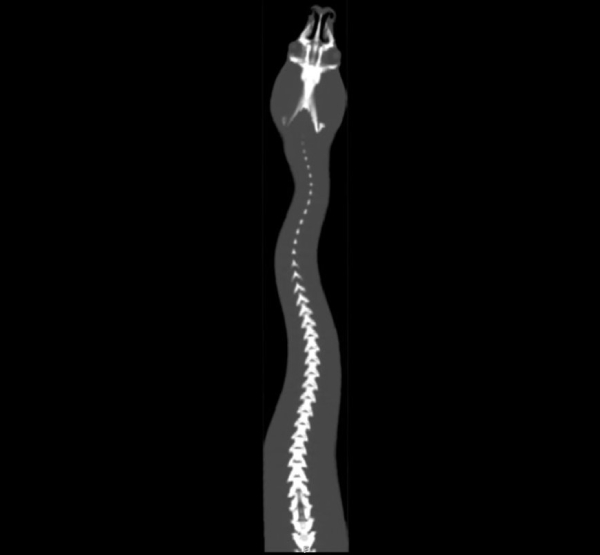
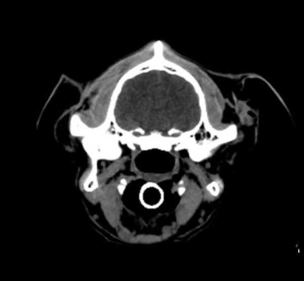
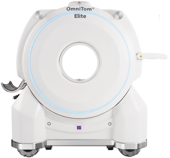
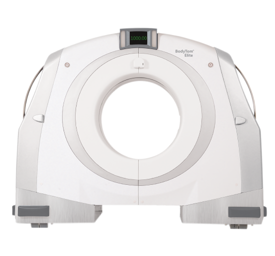
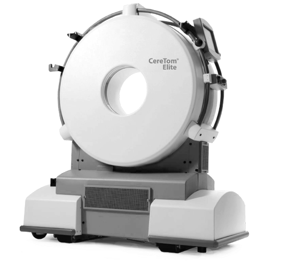
The NeuroLogica family of mobile CT scanners is battery-operated, charged from a wall outlet, and internally shielded. They provide diagnostic, multi-slice, and 3D imaging of soft tissue and bone.
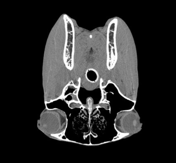
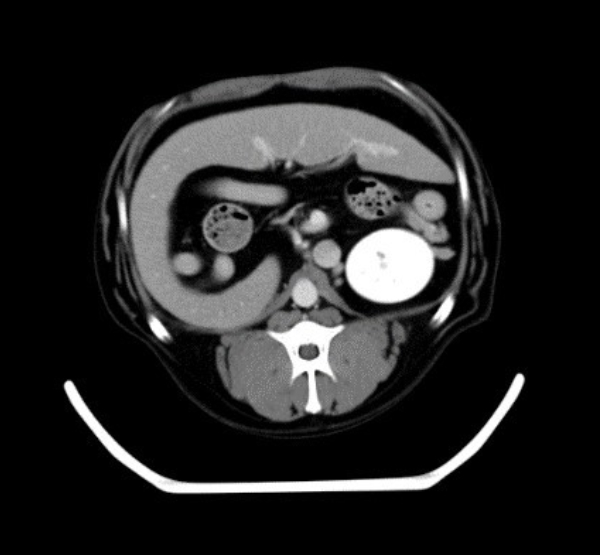
All our systems are multi-slice diagnostic CTs (not to be confused with Cone Beam CT or Flat Panel CT) that are utilized in human ICUs, ORs, Mobile Stroke Units, and other medical areas.
Treatment of cerebrovascular disease is a race against time, as it kills a large number of people worldwide and can cause paralysis in others. The main factor for reducing disability and death is a treatment 'time standard' within 270 minutes. These products save lives. Every minute counts.
Mount Sion Hospital
Everest, NY
Treatment of cerebrovascular disease is a race against time, as it kills a large number of people worldwide and can cause paralysis in others. The main factor for reducing disability and death is a treatment 'time standard' within 270 minutes. These products save lives. Every minute counts.
Dr. John Rolfstein
Veterinary Specialties Group, OH
Treatment of cerebrovascular disease is a race against time, as it kills a large number of people worldwide and can cause paralysis in others. The main factor for reducing disability and death is a treatment 'time standard' within 270 minutes. These products save lives. Every minute counts.
KU Medical Center
Kansas City, MO
Treatment of cerebrovascular disease is a race against time, as it kills a large number of people worldwide and can cause paralysis in others. The main factor for reducing disability and death is a treatment 'time standard' within 270 minutes. These products save lives. Every minute counts.
Mount Sion Hospital
Everest, NY
Treatment of cerebrovascular disease is a race against time, as it kills a large number of people worldwide and can cause paralysis in others. The main factor for reducing disability and death is a treatment 'time standard' within 270 minutes. These products save lives. Every minute counts.
Dr. John Rolfstein
Veterinary Specialties Group, OH
Treatment of cerebrovascular disease is a race against time, as it kills a large number of people worldwide and can cause paralysis in others. The main factor for reducing disability and death is a treatment 'time standard' within 270 minutes. These products save lives. Every minute counts.
Dr. John Rolfstein
Veterinary Specialties Group, OH
Dig Deeper
Read, watch and uncover the latest in mobile CT imaging
Foster, B., et al. (2014) “Segmentation of PET images for computer-aided functional quantification of tuberculosis in small animal models,” IEEE Transactions on Bio-Medical Engineering, 61(3), 711–724
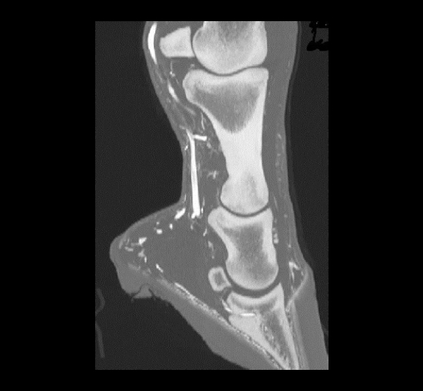
Gasiorowski, J.C., et al. (2015) “Clinical Use of Computed Tomography and Surface Markers to Assist Internal Fixation Within the Equine Hoof,” Veterinary Surgery, 44(2) 214–222
Porter, J.N., et al. (2014) “Altered cerebellar and prefrontal cortex function in rhesus monkeys that previously self-administered cocaine,” Psychopharmacology, 231(21), 4211-4218


.png?width=241&height=53&name=Case%20Studio%201%20(5).png)