The discovery of X-rays marked the beginning of a revolutionary change in the medical field and world. In today’s world, X-rays are used to diagnose several problems from broken bones to pneumonia to heart failure and much more… but not so long ago, these could only be discovered by cutting a person open. Wilhelm Konrad Roentgen, a Professor of Physics in Wurzburg, Bavaria discovered X-rays in 1895, accidentally! Roentgen was already known as an accomplished and dedicated scientist with very precise experimental methods. He was testing the effects of electron beams, “cathode rays” in electrical discharges through low-pressure gases. He uncovered that a screen coated with a fluorescent material placed outside of the discharge tube would project an incandescent glow even while shielded from the ultraviolet light of the discharge. He was able to show that there was an invisible radiation passing through the air to cause this fluorescence, and opaque objects placed between the tube and screen were transparent to the new form of radiation1. He discovered that this mysterious light could pass through the majority of substances but left shadows on solid objects. It is interesting to note, a few other scientists had come across this occurrence but none of them investigated it further, however Roentgen immediately realized he had stumbled upon something of high importance. He was unaware of what to name these rays so with “X” meaning unknown, he created the name “X-rays.” Quickly, he discovered that X-rays are able to pass through human tissue, making the bones as well as the tissue beneath visible.
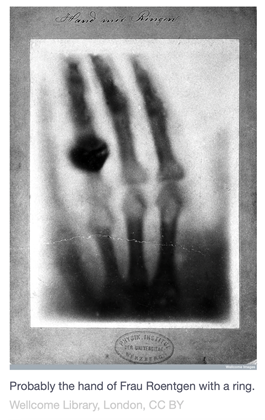
X-rays are electromagnetic energy waves that act similarly to light rays at wavelengths a thousand times shorter than those of light, they allow doctors to create images of internal organs and bones to help in diagnosing conditions or injuries. The defining characteristics of X-rays include their ability to penetrate opaque materials, the high energy of photons, and their wavelengths of atomic dimension. This discovery led to X-rays becoming one of the most important diagnostic tools in medicine, as it was the first way to see inside a human body without needing surgery. Before X-rays, doctors would normally treat on assumption depending on the symptoms the patient had, or had to perform surgery not knowing exactly what to look for. Broken bones, bullet locations, etc. were all diagnosed via physical examination and doctors’ best guess. Within two weeks of his discovery, he produced the first X-ray image of his wife Bertha’s hand. Almost immediately after this discovery, scientists and physicians started using the rays to look inside human bodies to reveal normal structures, bone fractures, kidney stones and foreign objects. In 1897, X-rays were used on a military battlefield for the first time during the Balkan War, to discover bullets and broken bones inside military patients. US President James Garfield actually died in 1881 due to not being able to locate the assassin’s bullet in his body, a century later thanks to X-rays, doctors were able to locate the bullet in President Reagan’s chest very quickly and saved his life.
When more research was conducted, later on a model of the atom was better explained for X-rays and there turned out to be two mechanisms for the production of X-rays. The first way X-rays are produced is the result of travelling electrons from the cathode and electrons in the glass atoms colliding. The second way X-rays are produced is the result of moving cathode electrons and penetrating deep inside the glas atom, hitting its nucleus. Both of these cases show that the result is not radiation that is composed of physical particles, like in the case of cathodic rays, but electromagnetic radiation that has more energy than visible light or radio waves.
Although scientists were able to quickly utilize the benefits of X-rays, they were slower to understand the harmful effects that x-ray radiation can cause for patients. Initially, people believed that X-rays passed through skin as harmlessly as light did, however, within several years researchers began to report cases of skin damage after being exposed to X-rays. In fact, during the 1930’s-1950’s, multiple American shoe stores actually featured shoe-fitting fluoroscopes using X-rays to allow customers to see the bones in their feet - just for fun, until the end of the 50’s when they realized this was very risky2. The high energy is what allows X-rays to deeply penetrate inside solid matter, X-rays are harmful because if an X-ray encounters a molecule while it’s traveling inside the body it could split that molecule open and shatter the chemical bonds that hold it together, which can cause real damage to tissue. In 1904, Thomas Edison began doing his own research on X-rays with a glassblower, Clarence Dally, who would X-ray his hands to test the tubes. Eventually, Dally had to have both of his arms amputated due to acute cancer and died of X-ray exposure. Dally’s death caused scientists to begin taking the risks of radiation more seriously, however it was still not fully understood. Later on Dr. D.W Gage of Nebraska discovered that there were dangers to radiation exposure such as reddened skin, skin lesions, and hair loss3.
Following the years after this discovery, we now have a far better understanding of the risks associated with X-ray radiation and there have been many protocols developed to help minimize unnecessary exposure. X-rays also have extended beyond just diagnosis, but investigators discovered that X-rays can be used to detect cancers in certain organs and treat some cancerous tumors, by directly affecting the DNA of cancerous cells. X-rays remain a keystone of modern medicine, newer medical imaging techniques relied on Roentgen’s discovery, such as computed tomography (CT), magnetic resonance imaging (MRI), ultrasound, echocardiography, and many others. In the 1970’s, computed tomography was developed, a new powerful X-ray imaging technique. Instead of X-rays being sent through the patient's body from one direction, beams of X-rays are sent from many angles, allowing it to be easier to create a sharp two-dimensional image of the interior of the body. CT scans are now playing an enormous role in medical diagnosis. A study showed that after a CT scan, the treating doctor’s leading diagnosis changes 42-51% of the time, sparing many patients from unnecessary surgeries and allowing for offering urgent treatment to patients faster than would otherwise be possible4.
The discovery of X-rays paved the way for the development of today’s broad amount of imaging techniques. What a great legacy for an accidental discovery!
Sources
1. https://www.britannica.com/science/X-ray/Fundamental-characteristics
2. https://www.history.com/this-day-in-history/german-scientist-discovers-x-rays
3. https://professionalradiologyep.com/the-history-of-x-rays-how-an-accidental-discovery-changed-the-world/
4. https://theconversation.com/on-the-120th-anniversary-of-the-x-ray-a-look-at-how-it-changed-our-view-of-the-world-50154