Early in the COVID-19 pandemic, doctors were faced with treating patients with severe lung infections, as well as neurological damages. A year into the pandemic, Dr. Abdelkader Mahammedi, M.D., assistant professor of radiology at UC, and Dr. Achala Vagal, M.D., professor of radiology, along with colleagues from Spain, Italy and Brazil, published a study based on 135 COVID-19 patients who had both respiratory and neurological complications. This showed that the patients with higher lung severity scores, were more likely to have acute abnormal neuroimaging findings.
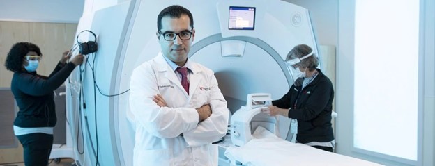
The CT imaging devices used to correlate the lung severity damage with the high risk of serious brain complications in COVID-19 patients, are powerful tools enabling physicians to provide the best therapies and care to those patients and improve outcome, including stroke prevention. CT imaging allows doctors to give better results and gain more confidence in the severity of illnesses and how they are formed.
Read the full article at Diagnostic Imaging