In essence, computed tomography (CT) are beams of x-rays rotating around a patient’s body that provide a detailed analysis of potential health issues, that cannot be identified by a simple x-ray. The first commercially available CT scanner was created in 1972 and the use of CT in veterinary medicine was first documented in the 1980’s to investigate diseases of the central nervous system and neoplasia in canines.1 Since then, the computed tomography has provided some of the greatest advancements in diagnostic imaging for the veterinary sector. In comparison to standard radiography, CT offers axial slices of the area observed, and allows a greater differentiation of soft tissue structures, and more importantly CT scanning limits scatter radiation; hence greatly enhancing images contrast. The most common areas of an animal's body that are imaged nowadays are the spine, nasal cavity, inner ear, bones/joints, and the chest/lungs. Due to technological advancements and increased availability in general practices, CT has become more and more common in veterinary medicine, along with reduced cost, improvements in availability and increase in the expertise and technology needed.
Many veterinary practices now have 8-slice to 64-slice CT scanners installed to treat animals of all shapes and sizes, not restricted only to small animal analysis and diagnosis. For example, Neurologica’s BodyTom, the world’s first battery-powered, portable, 32-slice CT scanner, was used to scan Layla, a 7-year-old, 2,300-pound eastern black rhinoceros at the Brookfield Zoo in Chicago.2 After being anesthetized and stabilized, Layla was slid onto a platform and gently moved onto a surgical table, where they performed the scan. As CT scanning is particularly valuable for imaging intracranial sections, as well as the nose, sinuses, and middle ear areas, which in Layla’s case, was critical to provide accurate diagnostic results and conclude that Laila needed sinus surgery. This diagnostic would have been very difficult to obtain and would have taken much longer and potentially involve more procedures, and very complex logistics due to Laila’s size and environment. Another example of large animal imaging procedures is related to the elephants. Unfortunately, elephants are very difficult to scan whilst alive, thus the focus has been on post-mortem tissues that veterinary practices have analyzed, which give valuable insights into pathologies. In elephants, there are many skeletal and foot problems, and with the use of CT scanning, the radiologists were able to provide bone modeling information, which is very valuable to the veterinarians. CT scanning studies of elephants’ foot bones showed that the damaged bone structure is mainly related to weight-bearing and aging, especially in zoos and safari parks environments. CT scanning has proven to be a great tool to provide information that will enable the animals’ caring management to adjust the environment and healthcare given to the animals so they can live longer lives with less discomfort. Studies using CT scanners have many beneficial outcomes for these animals, enhancing animal health and welfare in the long term.
The process for scanning an animal varies significantly depending on the animal size, but also the bones structures and soft tissues. The animals are moved by a specific number of millimeters, and this process is repeated to take images of the entire volume of interest without the imposition of structures. Modern multi slice CT scanners can acquire up to 620 images at once and can perform a complete scan of the abdomen or thorax in up to 10 seconds, image reconstruction time is short and compared to 25 years ago; the time of study is much faster to complete. In fact, in the mid 1980’s, to perform a CT scan on the skull of a small dog took more than an hour to acquire a dozen images, in low resolution. Today, using a 64-slice CT scanner, practices can perform a CT scan on skulls of smaller animals and generate 5,000 high quality images in a 10-to-15-minute examination. Even with these extremely fast systems, veterinary patients must be anesthetized and immobilized to perform the majority of these scans, but the period of anesthesia is short, and the value of information derived from the scans is extraordinary.
For small animals, CT is often used to treat patients with thoracic and abdominal diseases, intracranial and extracranial lesions, as well as any disorders of the musculoskeletal system (i.e., skeleton and spine.) Small animals are widely used for developing models in helping biomedical and biological research. In certain situations, as the CT scan is so rapid, this can be used in cases where anesthesia is not an option as it could compromise the patient. CT is also useful in emergency critical cases for small animals. In addition, there is the technical use of micro-computed tomography (μCT) in veterinary research/medicine for smaller animals. Micro-CT is similar to regular CT but on a smaller scale along with high resolution; some of the main advantages of Micro-CT are the high-spatial resolution, sensitivity to skeleton and lung, as well as the low-cost.
Another significant use of animal CT is in equine veterinary medicine, especially in diagnosing dental and sinus problems which causes head pain, and poor performance, which is a significant consideration within the horse racing training environment. To minimize the risk associated with a general anesthetic, some equine clinics have as setup where the horse stands on a platform and the horses head passes through the circular CT scanner.
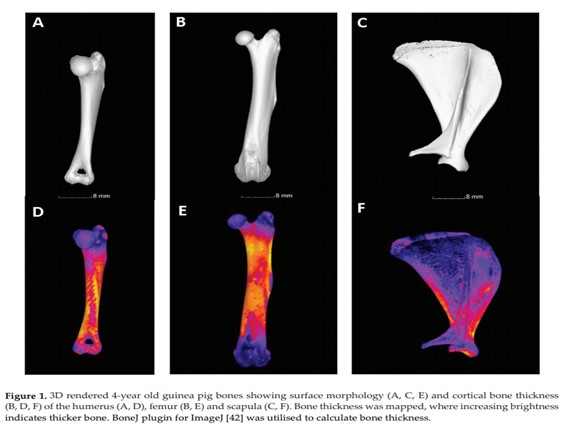
Growth and development are also observed using CT scanning in animals. There have been studies in human trabecular bone ontogeny, that showed an increase in trabecular bone thickness and bone volume fraction, but a decrease in trabecular number at around a year old, coinciding with the onset of unaided walking and load bearing was the cause of changes observed. Similar instances have been observed in cats, rats, and guinea pigs, in one study on a cat, they found that the bone density was analyzed and used to diagnose osteopenia, which is a condition that begins as you lose bone mass or bones get weaker, and the μCT was able to detect this and proper treatments were applied.
There is still a lot of research to be done for the use of CT scanner in veterinary medicine, further advancements in the clinic have been directed to using technology available alongside movement-restricting devices, to be able to create CT scan images without the need to euthanize a patient, thus decreasing the morbidity rates associated with general anesthesia. Equally as important in expanding the use of CT in veterinary medicine is the ability to create 3D reconstructions, this assists surgeons and medical personnel prior to the surgery and can be a valuable learning tool to teach more advanced anatomy and physiology of animals.
References
1 https://www.intechopen.com/books/computed-tomography-advanced-applications/computed-tomography-in-veterinary-medicine-currently-published-and-tomorrow-s-vision
2 https://www.czs.org/LaylaRhino