A commonly asked questions is, "what is the difference between a PET Scan and a CT scan?"
CT Scan
A Computed Tomography, otherwise known as a CT scan, uses computers and rotating X-ray machines to create cross-sectional images of the body. These images provide more detailed information than normal X-ray images. For example, they can show the soft tissues, blood vessels, and bones in various parts of the body, such as the head, shoulders, spine heart, abdomen, knee, and chest.
During a CT scan, you lie in a tunnel-like machine while the inside of the machine rotates and takes a series of X-rays from different angles. These pictures are then sent to a computer, where they are combined to create images of slices, or cross-sections, of the body. They may also be combined to produce a 3-D image of a particular area of the body. The test itself is minimally invasive and can be conducted quickly – anywhere from 20 minutes to one hour.1
There are many uses for a CT scan, but it’s particularly well-suited for diagnosing diseases and evaluating injuries. The imaging technique can help your doctor with the following:
- diagnose infections, muscle disorders, and bone fractures
- pinpoint the location of masses and tumors (including cancer)
- study the blood vessels and other internal structures
- assess the extent of internal injuries and internal bleeding
- guide procedures, such as surgeries and biopsies
- monitor the effectiveness of treatments for certain medical conditions, including cancer and heart disease
Doctors may give the patient a special dye called a contrast material to help internal structures show up more clearly on the X-ray images. The contrast material blocks X-rays and appears white on the images, allowing it to highlight the intestines, blood vessels, or other structures in the area being examined. Depending on the part of the body being inspected, the patient may need to drink a liquid containing the contrast. Alternatively, the contrast may need to be injected into the patient’s arm or ad-ministered through their rectum via an enema.
There are very few risks associated with a CT scan. Though CT scans expose patients to more radiation than typical X-rays, the risk of cancer caused by radiation is very small if there is only have one scan. The risk for cancer may increase over time if there are multiple X-rays or CT scans. It should be noted, the risk of cancer is increased in children receiving CT scans, especially to the chest and abdomen.
PET Scan
A positron emission tomography, or PET scan is an imaging test that allows doctors to check for diseases in the body.
The scan uses a special dye containing radioactive tracers. These tracers are either swallowed, inhaled, or injected into a vein in the patient’s arm depending on what part of the body is being examined. Certain organs and tissues then absorb the tracer. When detected by a PET scanner, the tracers help doc-tors see how well the patient’s organs and tissues are working.2
The tracer will collect in areas of higher chemical activity, which is helpful because certain tissues of the body, and certain diseases, have a higher level of chemical activity. These areas of disease will show up as bright spots on the PET scan.
A PET scan is typically an outpatient procedure.
Doctor’s may order a PET scan to inspect a patient’s blood flow, oxygen intake, or the metabolism of organs and tissues. PET scans show problems at the cellular level, giving doctors the best view of com-plex systemic diseases.
PET scans are most commonly used to detect the following:
- Cancer
- Heart problems
- Brain disorders, including problems with the central nervous system (CNS)
Importantly, cancer cells have a higher metabolic rate than noncancerous cells. Because of this high level of chemical activity, cancer cells show up as bright spots on PET scans. For this reason, PET scans are useful both for detecting cancer and for:
- seeing if the cancer has spread
- seeing if a cancer treatment is working
- checking for a cancer recurrence
With regard to heart problems, PET scans reveal areas of decreased blood flow in the heart. This is be-cause healthy heart tissue will take in more of the tracer than unhealthy tissue or tissue that has de-creased blood flow.
Different colors and degrees of brightness on the scan will indicate different levels of tissue function, helping the patient and doctor decide how best to move forward.
And regarding brain disorders, glucose is the main fuel of the brain. During PET scans, tracers are “attached” to compounds such as glucose. By detecting radioactive glucose, the PET scan is able to detect which areas of the brain are utilizing glucose at the highest rates. A doctor will look at the scan to see how the brain is working and to check for any abnormalities.
PET scans are also used to help diagnose and manage many central nervous system (CNS) disorders, including:
- Alzheimer’s disease
- Depression
- Epilepsy
- Head Trauma
- Parkinson’s disease
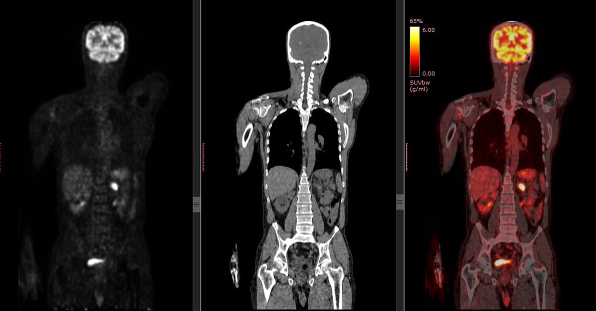
CT and PET Scan Comparison
PET scans show metabolic changes occurring at the cellular level in an organ or tissue. This is important because disease often begins at the cellular level. CT scans (and MRIs) can’t reveal problems at the cellular level.
PET scans can detect very early changes in a patient’s cells. CT scans (and MRIs) can only detect changes later, as a disease alters the structure of your organs or tissues.
Detection of illness at the cellular level gives medical professionals the best view of complex systemic diseases.
Perhaps the main difference between a CT scan and a PET scan is their focus. A CT scan creates a de-tailed non-moving image of organs, bones, and tissues. A PET scan, on the other hand, shows doctors how the tissues in a patient’s body work on a cellular level. Other important differences include:
- They use different materials: CT scans pass x-rays through the body to create images. A PET scan uses a radioactive material which emits energy. The energy is then detected by a special camera to produce images.
- A PET scan is more time-consuming: A CT scan is performed in minutes. This makes it an excel-lent tool in emergency situations when doctors need to act fast. A PET scan could take 20 minutes to several hours, depending on a patient’s condition. In some cases, the procedure may be performed over several days.
- Radiation does not stay in the body with a CT scan: After a CT scan, no radiation remains in the body. On the contrary, a small amount of radiation may stay in the body for a short time after a PET scan.
- PET scans detect cancer earlier than other tests: Unlike other forms of imaging, a PET scan shows molecular activity and helps doctors identify diseases in the earliest stages. For this rea-son, a PET scan is a highly reliable tool for detecting cancer. CT scans show signs of an issue after a disease begins to change the structure of tissues or organs.
References
1. Cassoobhoy, A. (2020, December 13). CT scan (CAT Scan): Purpose, Procedure, risks, Side-Effects, Results. Retrieved April 25, 2021, from https://www.webmd.com/cancer/what-is-a-ct-scan
2. Kiefer, D. (2018, June 06). Lung pet Scan: Purpose, procedure, and preparation. Retrieved April 25, 2021, from https://www.healthline.com/health/lung-pet-scan