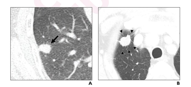
Figure. Axial contrast-enhanced CT images in patients with and without the presence of pathologic lymphovascular invasion.
This article from the American Journal of Roentgenology includes a retrospective study including 900 patients with stage IA non-small cell lung cancer. By using features of computed Tomography (CT), the study focuses on identifying patients who are good candidates for sublobar resection instead of undergoing more extensive surgery. Currently, lobectomy with mediastinal lymph node dissection is the treatment for IA non-small cell lung cancer, however less invasive treatment options are preferable especially given the pulmonary comorbidities in the older population.
Through chest CT examinations, using 64-slice CT scanners; doctors were able to evaluate the prognostic values of the CT features, in patients undergoing sublobar resection, and gather information on patients with the presence of pathologic lymphovascular invasion (pLVI.). CT scanners have the ability to improve treatments like this, and allow for less invasive and less harmful radiation to patients in the future, and provide efficient diagnostic performance.
Read the full article at the American Journal of Roentgenology.