Computed Tomography, or CT, refers to computerized imaging procedures where an x-ray beam is aimed towards the patient and rotated around the body, in order to create cross-sectional images of the body. The word “tomography” is derived from Greek for, “tomos” meaning section or slice and “graphe” meaning drawing. In referring to CT scanners, the word “slice” is often mentioned, but does it refer to?
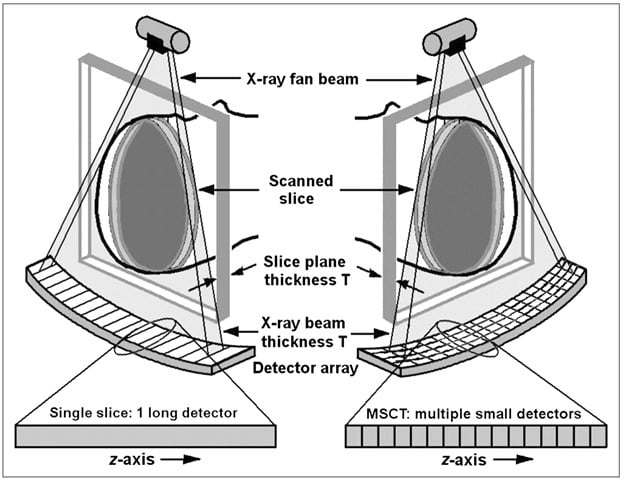
The term slice refers to the number of rows of detectors in the z-axis of a CT. For example, in an 8-slice CT, there are eight slices of data captured for each rotation of the gantry. The first CT scanners offered single slice CT (SSCT) images but now there are multiple-slice CT scanners (MSCT.) The limitation with using a SSCT was that the thinner slices requiring high image-quality were not achievable unless the region to be scanned was very restricted, leading to low-quality images. A solution to this issue was to utilize the x-ray beam, incorporating multiple rows of detectors, thereby collecting more than one slice at a time and reducing the number of rotations needed. This method also led to the development of MSCT technology. The primary difference in the hardware between the two methods is the design of the detector arrays. SSCT detector arrays are one dimensional, consisting of high numbers of detector elements in a single row, whereas the MSCT allows for each individual element to be divided into several smaller detector elements creating a 2-dimensional array. As seen in the image, as opposed to a singular row of detectors along the fan beam, there are multiple rows of detectors.
The first scanner with more than one row of detectors was introduced by Elscint in 1992 and was called the CT-Twin. This scanner allowed data for 2 slices to be shown simultaneously; this addressed x-ray heating problems, and significantly reduced scanning time. Eventually, the first “modern” versions of MSCT scanners were developed and introduced in 1998 and simultaneously acquired 4 slices, which meant four detector rows corresponding to four data channels. In 2002, the first MSCT scanners providing16 slices were introduced.
In the current market, the commonly available CT slice counts include 16, 32, 40, 64, and 128 slices, with less common ones providing up to 256 and 320 slice CT scanners. The 4 to 8 slice scanners are slowly being withdrawn from the market.
When patients are put through the CT, the circular opening rotates to take a series of x-rays with each rotation taking approximately 1 second. Multiple slice CT scanners initially could take four separate images through each rotation, but technology has improved to the level that CT scanners can now take between 6 to 128 separate images in a singular rotation, meaning that it takes significantly less time to complete a CT scan.
Different slice-counts for CT scans can be useful for many different scenarios. The majority of CT scanners can perform general imaging procedures, to include chest and head exams as well as multiple different body views to scan for any fractures. However, for cardiac procedures, higher slice counts are required to ensure optimum image quality. Multi-slice CT scanners have numerous advantages such as superior image quality and this can enable earlier diagnostic results. This essentially leads to shortening the diagnostic time for the patient, enhancing the treatment, and improving the patients’ long-term outcome. Radiation dosage is always a major concern when getting a CT scan, and with the higher slice CT systems, there is the additional benefit of reducing this dose. With technologies such as automatic exposure control (AEC) and iterative reconstruction (IR), a patient scanned on a higher slice CT will receive significantly lower doses of radiation than a patient on a lower dose CT scanner. MSCT can improve overall patient experience as well.
CT scanning is an inherently unsettling experience and now with the ability to capture images faster with multiple slice scanners, patients are able to spend less time on the table, and the scanner puts the images into physicians’ hands faster. MSCT also allows large anatomic body ranges to be scanned producing thin and thick sleeves; thick slices are important for primary interpretation, and thin slices are important for reducing partial-volume streaks and allowing for high quality 3-dimensional reconstructions. In summary, the higher the slice count, the faster the speed of the scan; a conventional single-slice CT scanner may take up to ten minutes to complete a scan whereas multi-slice scanners are able to do the job within seconds. Shortening the time for scanning is especially useful for the treatment of children or others who may find it difficult to lie in one position for an extended period of time.
16-slice CT scanners are the ideal machines for higher-use facilities and for everyday use, particularly where reducing scan time is important. It is a good fit for Urgent Care Centers and hospitals alike. However, 32 and 64 slice CT scanners are becoming standard for imaging centers and hospitals; the accuracy and speed make them very suitable for hospitals with higher patient throughput. These higher slice CT scanners provide longer coverage per gantry rotation than the 16 slice scanners and reduce the likelihood of motion artifacts, which can cause blurring or double images in scans. The BodyTom Elite from Neurologica, a subsidiary of Samsung Electronics, is the world’s first mobile, full-body, 32-slice CT scanner, which incorporates the higher CT slice count as well as the portable aspect which allows for transporting the machine right to the patient’s bedsides for any procedures, as opposed to the traditional method of transporting patients to the radiology room. With the two combined, systems like this can save facilities valuable time and money.
In conclusion, there are many advantages to the multiple slice CT scanners over the single slice scanners; these machines can increase the diagnostic capabilities of the scan, resulting in clearer images for the medical professionals, a diminished exposure of radiation for the patients, and better long-term outcomes. Multiple slice CT scanners will continue to evolve and grow as they have become a primary diagnostic imaging tool.